
Atharva Shetye
I am a Ph.D. researcher in Mechanical Engineering at UCLA and former R&D Design Engineer at BD, with experience spanning medical device development, mechanical design, computational mechanics, and biomedical imaging. My work combines CAD, FEA, experimental mechanics, and advanced image analysis methods to study complex mechanical behavior in biological systems and develop engineering solutions for medical applications.
At BD, I led mechanical design activities across multiple medical device product lines, translating requirements into manufacturable designs through CAD, GD&T, tolerance stack-up analysis, FEA, DFM, testing, and cross functional product development. My doctoral research builds on this engineering foundation through computational modeling, experimental characterization, and image based measurement of ocular tissue mechanics.
I am a named inventor on 8 U.S. patent applications and author of 2 peer reviewed publications. This portfolio highlights selected projects across mechanical design, simulation, experimental mechanics, and computational imaging.
Work & R&D Experience

Doctoral Researcher @ Jules Stein Eye Institute, UCLA
Los Angeles, CA
PhD Research Projects:

R&D Design Engineer II @ Becton Dickinson and Company (BD)
Bengaluru, India
BD Key R&D Project:
Featured Projects
3D CAD and Dynamic FEA of Ocular Mechanics During Saccades
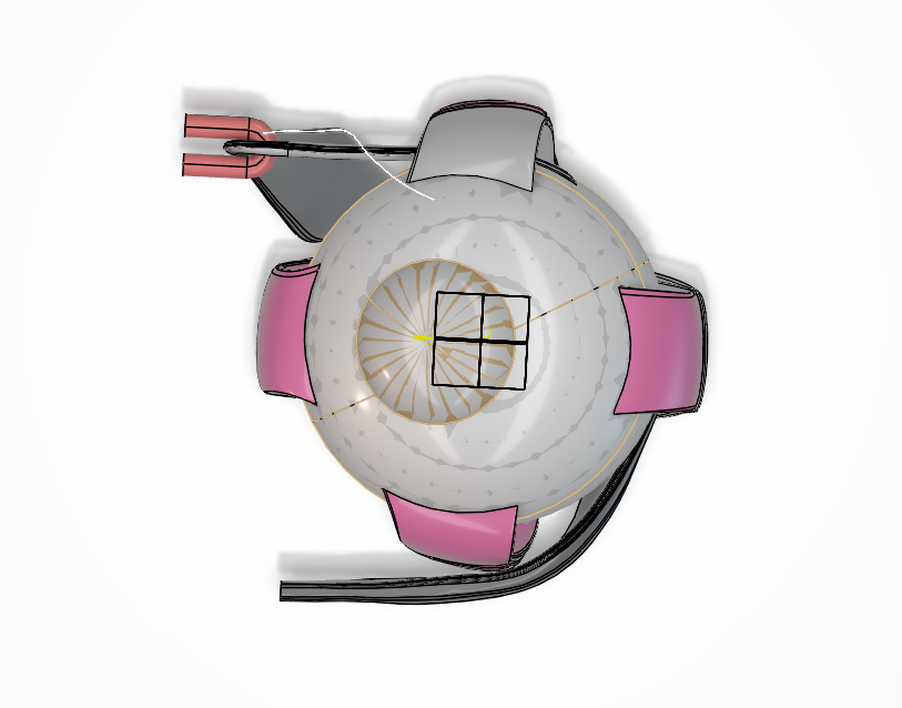
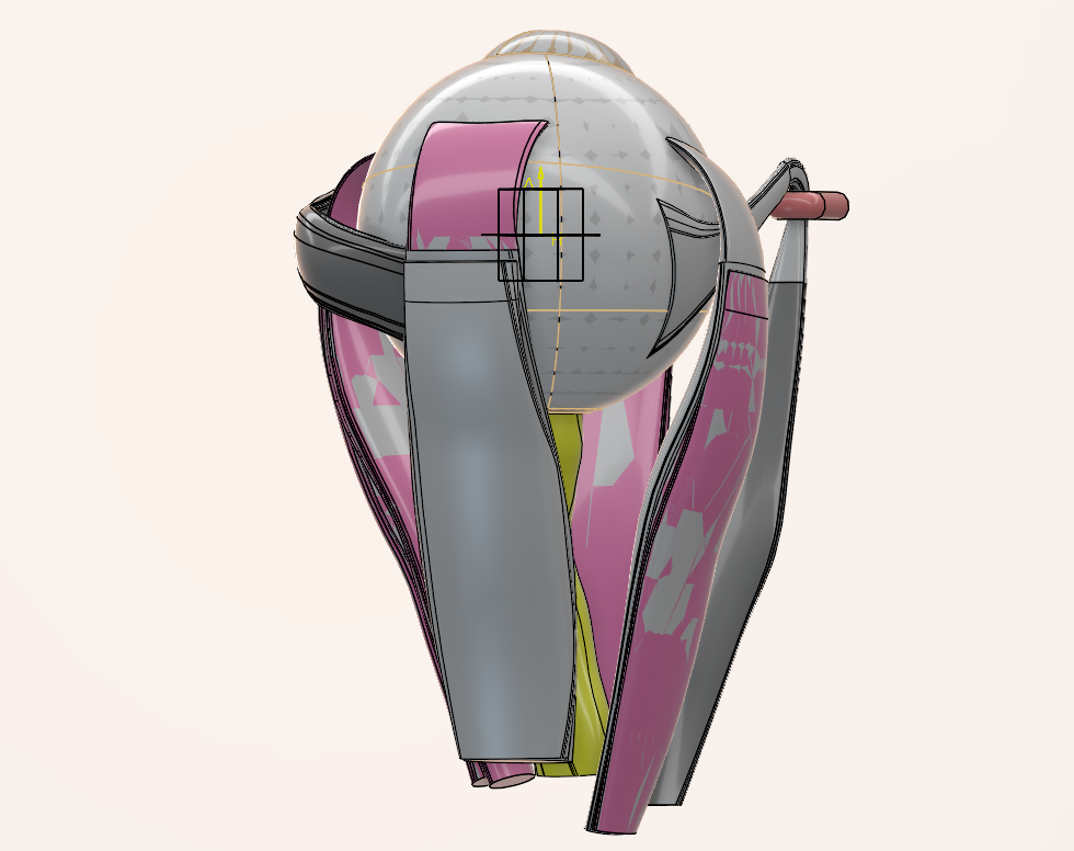
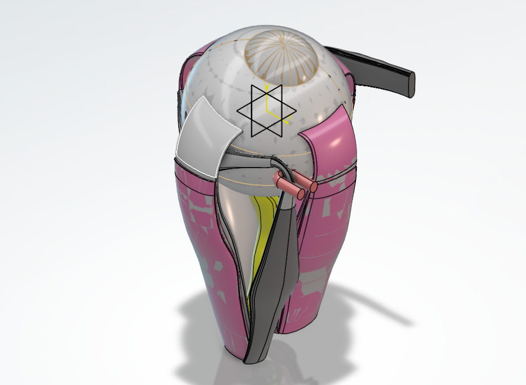
Developing an anatomically consistent 3D ocular model in CATIA and Abaqus to simulate extraocular muscle activation and quantify transient stress, strain, and deformation during rapid eye movements.
Viscoelastic Material Characterization of Human Ocular Tissues
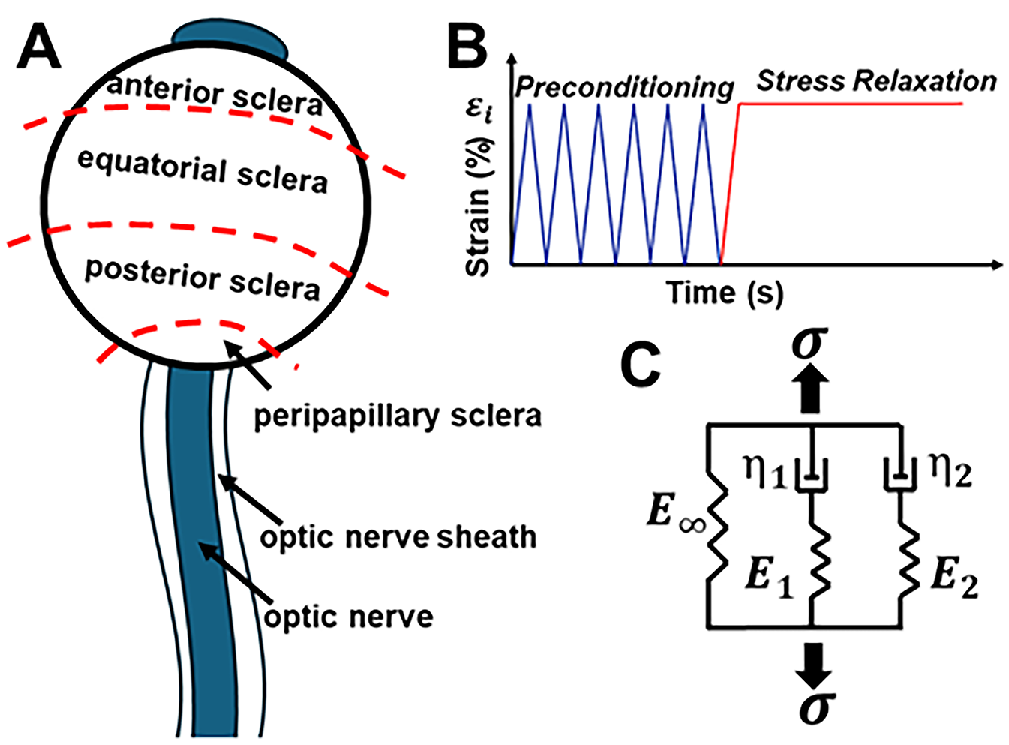
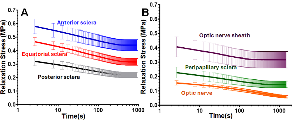
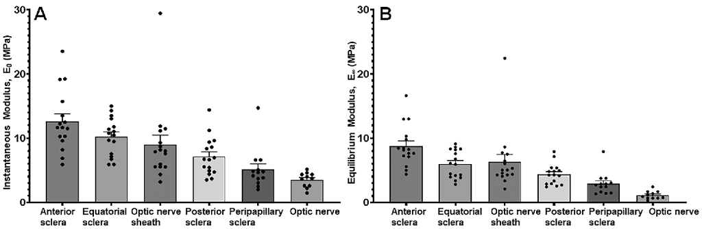
Characterized 6 human ocular tissue regions through tensile stress relaxation testing and generalized Maxwell/Prony-series modeling to derive time dependent material properties.
In Vivo Measurement of Gaze Induced Ocular Strain
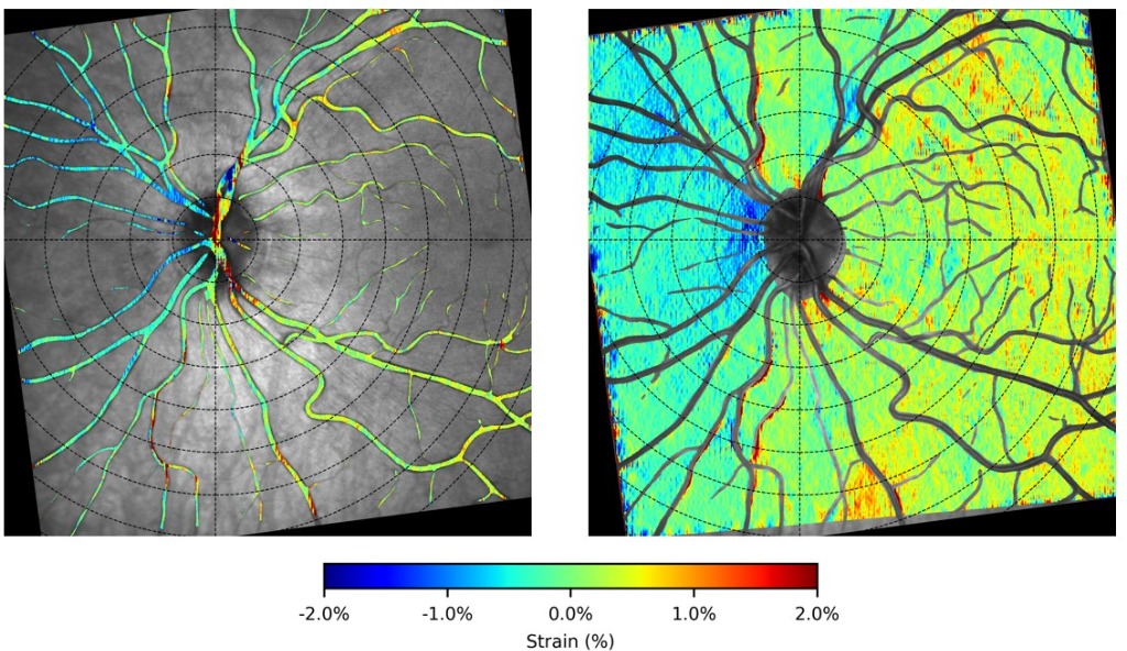
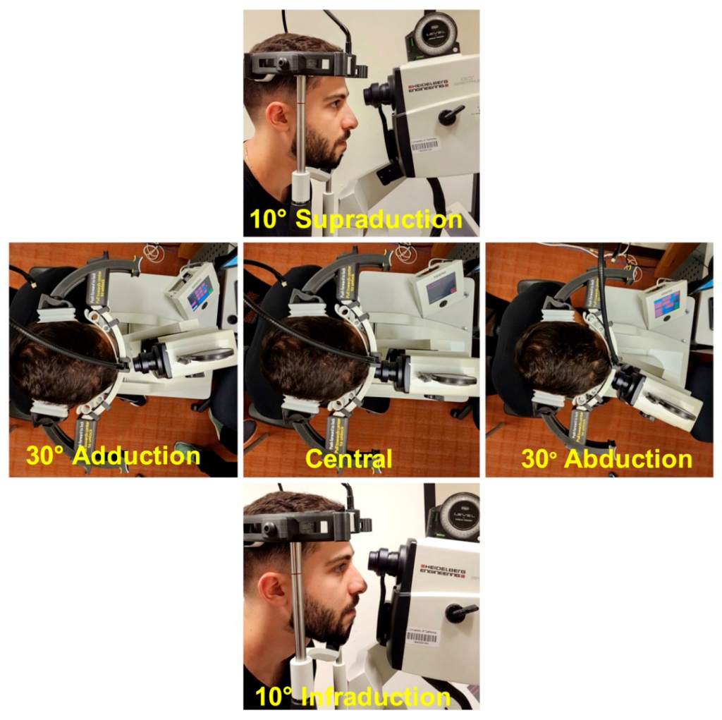
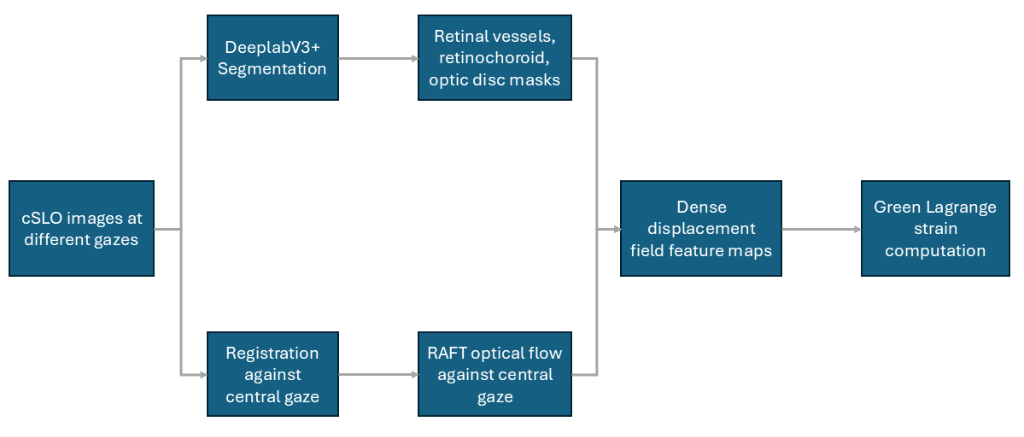
Developed an image based mechanics pipeline combining deep learning segmentation, image registration, RAFT optical flow, and continuum mechanics to quantify full field ocular strain during gaze shifts.
Cardiac Pulse Estimation from Retinal Strain in Sequential cSLO Imaging
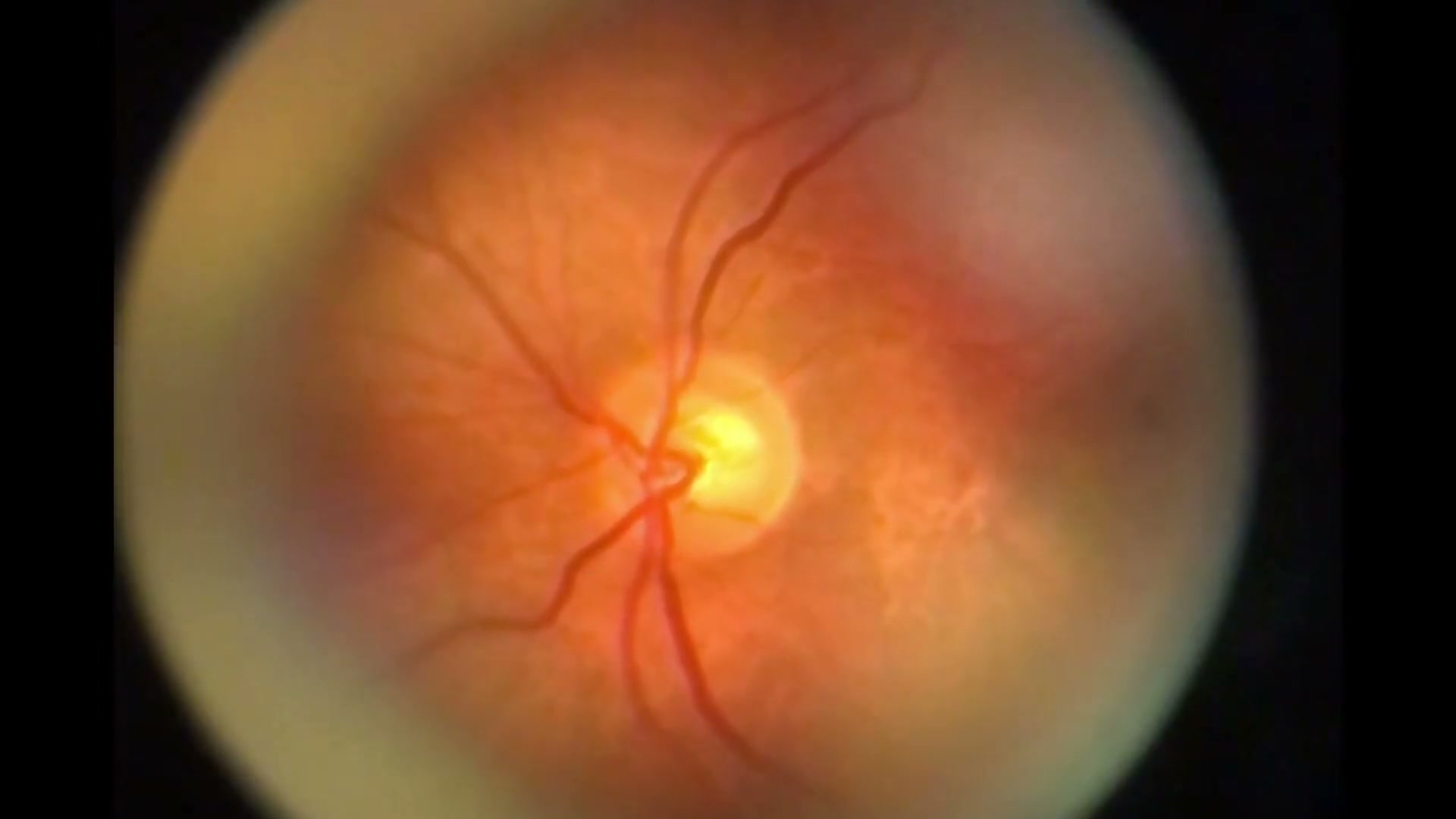
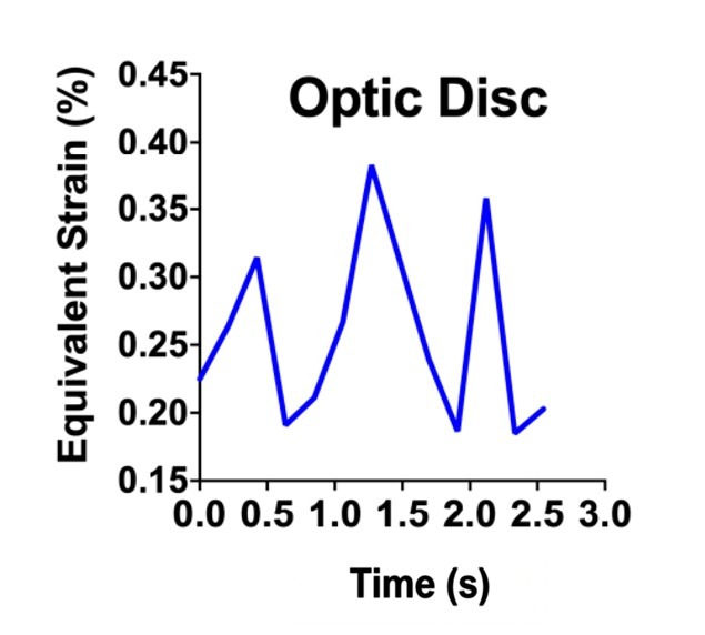
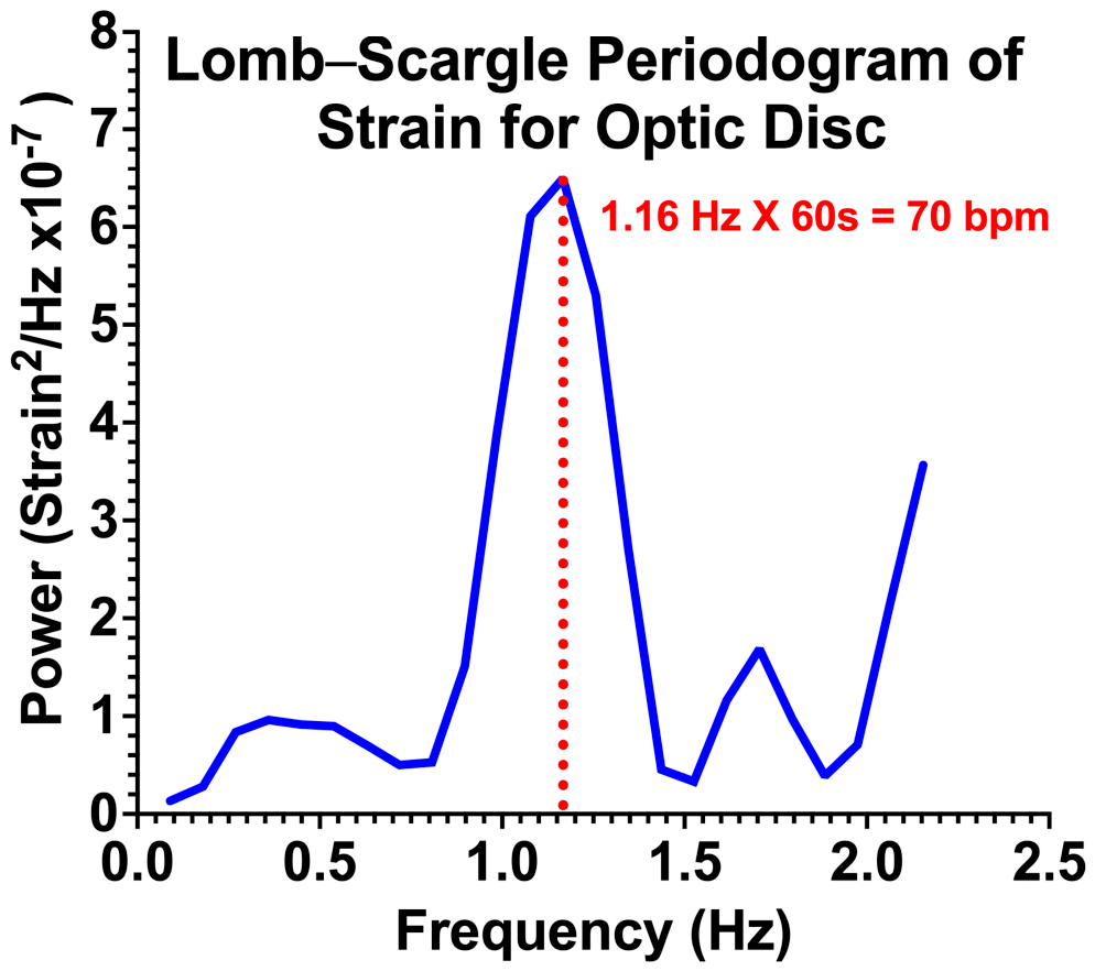
Quantified time varying retinal vessel strain from sequential cSLO imaging and used FFT and Lomb–Scargle spectral analysis to identify cardiac pulse related deformation frequencies.
In Vivo Estimation of Retinal Viscoelastic Relaxation from High-Speed cSLO
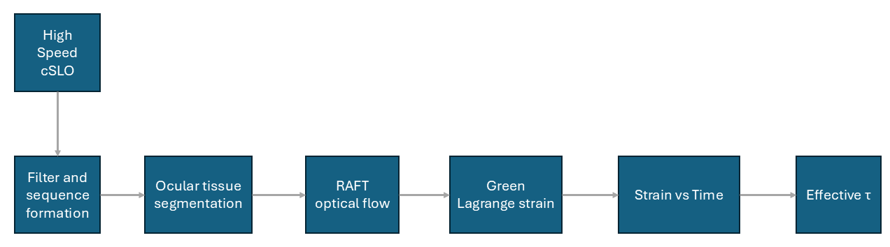
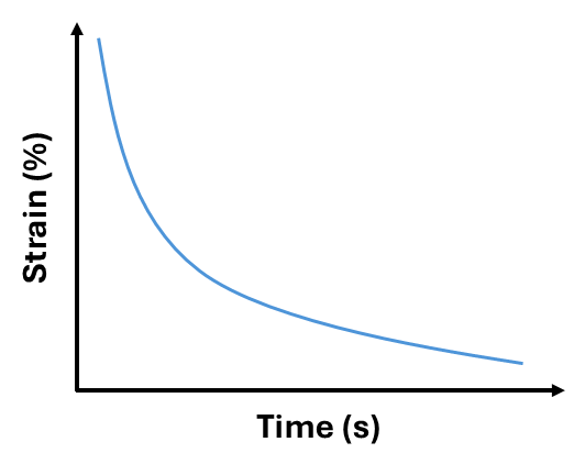
Developing a high-speed image analysis workflow to track post saccadic retinal strain decay and non invasively estimate effective viscoelastic relaxation time constants in vivo.
BD Hypodermic Luer Redesign & ISO 80369-7 Transition
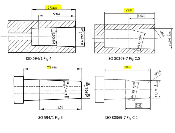
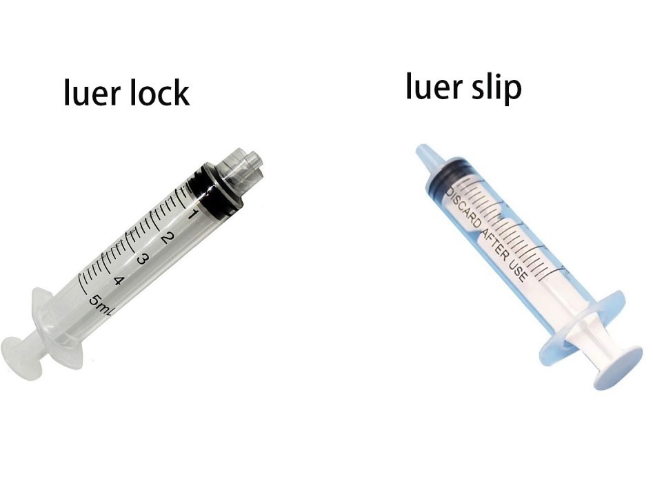
Redesigned Luer lock and Luer slip interfaces across 9 high volume product lines using SolidWorks, GD&T, tolerance analysis, Abaqus FEA, and DFM while minimizing manufacturing and tooling changes.
Education & Research Training
Ph.D. in Mechanical Engineering
University of California, Los Angeles (UCLA)
Los Angeles, CA•GPA 3.90 / 4.00
Focused on studying the mechanical response of ocular tissues during static gaze fixations and dynamic eye movements. Using principles of mechanical engineering, currently working towards tissue property characterization using deep learning based image analysis, dynamic FEA, and experimental tests to quantify real time ocular tissue response under physiological dynamics.
M.S. in Mechanical Engineering
University of California, Los Angeles (UCLA)
Los Angeles, CA•GPA 3.90 / 4.00
Specialized in the design of mechanical systems and robotic manipulators. Coursework included compliant mechanisms, and kinematics, dynamics, and control of robotic arms.

B.Tech. in Production Engineering
National Institute of Technology, Tiruchirappalli
Tiruchirappalli, India•GPA 3.93 / 4.00
Core production and industrial engineering program focusing on advanced manufacturing processes, CAD/CAM prototyping, operations research, quality control, precision machining, and industrial automation.
Journal Publications
Optic Disc and Retinal Strain Due to Vertical Duction
Investigative Ophthalmology & Visual Science (IOVS, September 2026)
Confocal scanning laser ophthalmoscopy (cSLO) and image-based strain analysis were used to evaluate optic disc and peripapillary retinal vessel deformation during vertical versus horizontal ductions. Notably, just 10° of vertical rotation produced strain magnitudes comparable to 30° of horizontal duction, with strain peaking in the optic disc and younger adults exhibiting approximately twofold higher strain than older adults, demonstrating significant optic nerve tethering during vertical gaze shifts.
Evidence That Cardiac Pulse Strains Retinal Vessels in and near the Optic Disc During Ocular Ductions
Bioengineering Journal (MDPI, June 2026)
Sequential confocal scanning laser ophthalmoscopy (cSLO) images were analyzed to quantify cardiac pulse induced retinal vessel strain and evaluate its variation across different gaze positions. Deep learning segmentation, RAFT optical flow, Green–Lagrangian strain analysis, and spectral methods identified pulse related deformation, with mean equivalent strain ranging from approximately 0.27% to 0.36%, exceeding the 0.16% noise threshold across gaze positions.
Linear Viscoelasticity of Human Ocular Tissues During Tensile Stress Relaxation
Investigative Ophthalmology & Visual Science (IOVS, May 2026)
Tensile stress relaxation testing was used to characterize the viscoelastic behavior of six human ocular tissue regions and derive region specific material properties for computational modeling. Generalized Maxwell models represented through Prony-series parameters quantified substantial regional differences in stiffness and relaxation behavior, with instantaneous modulus ranging from approximately 3.5 MPa in the optic nerve to 12.6 MPa in the anterior sclera and long term relaxation times ranging from approximately 60 to 460 seconds.
Patents & Inventions
Disinfecting Cap for Male and Female Needleless Connectors with Clamping Arms
U.S. Patent Application US20240269454A1
Disinfecting Cap for Male and Female Connectors Including Deformable Housing
U.S. Patent Application US20240245899A1
Medical Device Kit with Integrated Organization and Waste Disposal Features
U.S. Patent Application US20240139466A1
Universal Disinfection Device with Rotatable Sleeve
International Patent Application WO2025198953A1
Universal Fluid Connector Assembly with a Regulating Valve Component
U.S. Patent Application US20240123211A1
Secondary Securement Device for Vascular Access Device I
U.S. Patent Application US20250082904A1
Secondary Securement Device for Vascular Access Device II
U.S. Patent Application US20240424261A1
Secondary Securement Device for Vascular Access Device III
International Patent Application WO2024151251A1
Awards & Honors
Graduate Student Research Excellence Award
Jules Stein Eye Institute · UCLA
Awarded for excellence in biomechanics research, specifically for developing deep learning segmentation and optical flow pipelines to quantify ocular tissue strain and deformation during eye movements.

Retina Research Foundation Travel Grant
Association for Research in Vision and Ophthalmology (ARVO)
Competitive travel grant awarded to present novel research on ocular tissue strain in peripapillary retina due to eye movements at the Association for Research in Vision and Ophthalmology (ARVO) annual conference.
BD "Drive Results" Award for Design Excellence (FY23)
Becton Dickinson (BD)
Awarded for executing key design deliverables related to product launch.
BD "Strong Teams" Award for Cross-Functional Leadership
Becton Dickinson (BD)
Awarded for driving essential cross-functional collaboration to deliver core fiscal-year business objectives.